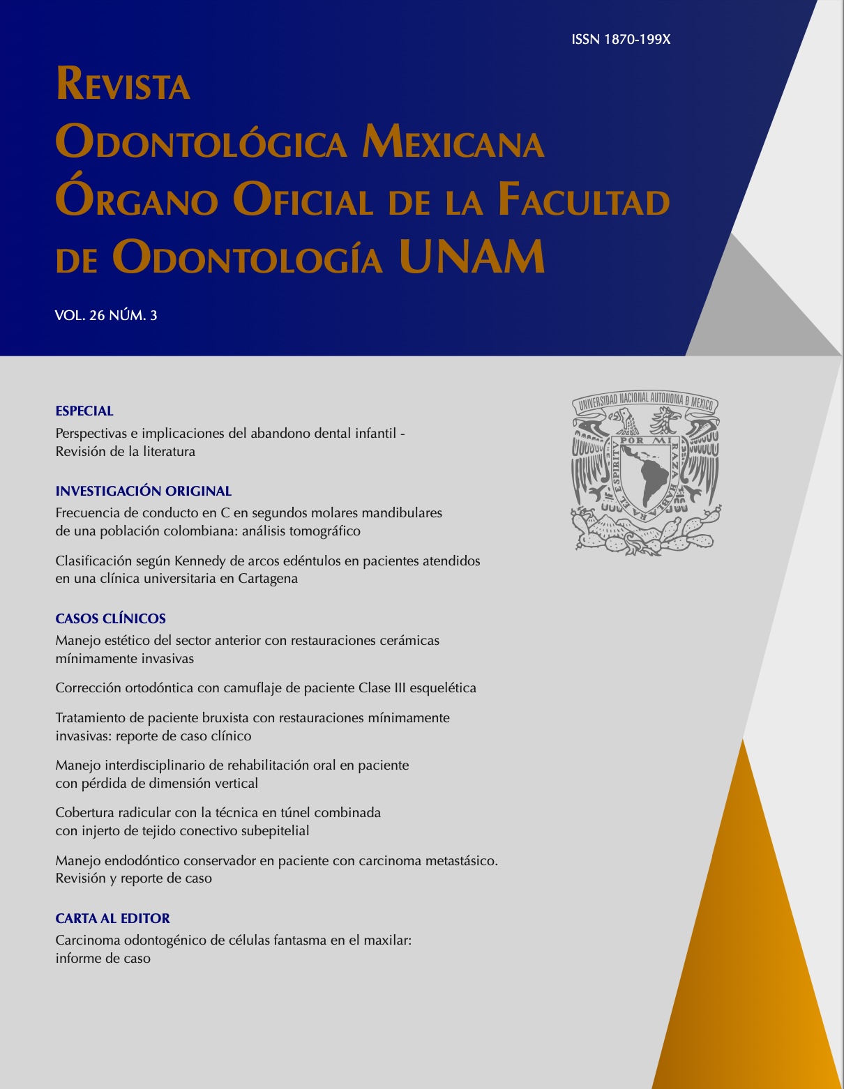
Frecuencia de conducto en C en segundos molares mandibulares de una población colombiana: análisis tomográfico
Contenido principal del artículo
Resumen
Introducción: La configuración del canal radicular en forma de C es una variación anatómica común en segundos molares mandibulares con fusión radicular, con una prevalencia de entre 2.7% y 44.5%. Objetivo: Estimar la frecuencia de conductos radiculares en forma de C en segundos molares mandibulares por medio de tomografías computarizadas tomadas a pacientes que acudieron a centros radiológicos. Materiales y métodos: Se evaluaron 120 segundos molares mandibulares. Se determinó la frecuencia y tipo de conducto en C de acuerdo con el género, y ubicación de la pieza dentaria. Se usó el software Invivo 5. Para el análisis estadístico de datos (p=0.05), se realizó la prueba de Chi-cuadrada. La forma de conducto en C se categorizó con base en la clasificación de Fan y la presencia de ranura y surco con base en la clasificación de Shemesh et al. 2017. Resultados: La frecuencia de conductos en forma de C en la población estudiada fue de 25%, encontrando diferencia significativa en cuanto al género. La forma del conducto en C a nivel de los tercios radiculares cervical y apical fue más frecuente para el tipo II y en medio para el tipo III. Se encontró que 6.7% de la población estudiada presentaban tipo I según la clasificación de Shemesh et al. 2017, con más frecuencia en el lado derecho (62.5%), mientras que 3.4% presentaban tipo III con mayor frecuencia en el lado izquierdo (75%). Conclusiones: Este es uno de los pocos estudios realizados en Colombia relacionados con la frecuencia del conducto en C y se logró determinar que hay mayor frecuencia en el género femenino (33.3%).
Detalles del artículo
Citas en Dimensions Service
Citas
Cooke HG, Cox FL. C-shaped canal configurations in mandibular molars. J Am Dent Assoc. 1979; 99(5): 836-839. DOI: 10.14219/jada.archive.1979.0402
Sherwood IA, Gutmann JL, Kumar S, Evangelin J, Nivedha V, Sadashivam V. CBCT analysis of the anatomy of C-shaped root canals in mandibular second molars from a southern Indian population in Tamil Nadu. Endodontic Practice Today. 2019; 13(1): 61–70
Shemesh A, Levin A, Katzenell V, Itzhak JB, Levinson O, Avraham Z, et al. C-shaped canals —prevalence and root canal configuration by cone beam computed tomography evaluation in first and second mandibular molars— a cross-sectional study. Clin Oral Investig. 2017; 21(6): 2039-2044. DOI: 10.1007/s00784-016-1993-y
Ren HY, Zhao YS, Yoo YJ, Zhang XW, Fang H, Wang F, et al. Mandibular molar C-shaped root canals in 5th millennium BC China. Arch Oral Biol. 2020;117: 104773. DOI. 10.1016/j.archoralbio.2020.104773
Jafarzadeh H, Wu YN. The C-shaped root canal configuration : A review. J Endod. 2007; 33(5): 517-523. DOI: 10.1016/j.joen.2007.01.005
Melton DC, Krell KV, Fuller MW. Anatomical and histological features of C-shaped canals in mandibular second molars. J Endod. 1991; 17(8): 384-388. DOI: 10.1016/S0099-2399(06)81990-4
Fan B, Cheung GSP, Fan M, Gutmann JL, Bian Z. C-shaped canal system in mandibular second molars : Part I — anatomical features. J Endod. 2004; 30(12): 899-903. DOI: 10.1097/01.don.0000136207.12204.e4
Martins JNR, Mata A, Marques D, Caramês J. Prevalence of root fusions and main root canal merging in human upper and lower molars : A cone-beam computed tomography in vivo study. J Endod. 2016; 42(6): 900-908. DOI: 10.1016/j.joen.2016.03.005
Martins JNR, Marques D, Silva EJNL, Caramês J, Mata A, Versiani MA. Prevalence of C-shaped canal morphology using cone beam computed tomography – a systematic review with meta-analysis. Int Endod J. 2019; 52(11): 1556-1572. DOI: 10.1111/iej.13169
Quijano S, García C, Rios K, Ruiz V, Ruíz A. Sistema de conducto radicular en forma de C en segundas molares mandibulares evaluados por tomografía cone beam. Rev Estomatol Herediana. 2016; 26(1): 28-36. http://www.scielo.org.pe/scielo.php?script=sci_arttext&pid=S1019-43552016000100005
Kato A, Ziegler A, Higuchi N, Nakata K NH, Nakamura H, Ohno N. Aetiology, incidence and morphology of the C shaped root canal system and its impact on clinical endodontics. Int Endod J. 2014; 47(11): 1012-1033. DOI: 10.1111/iej.12256
Silva EJNL, Nejaim Y, Silva AV, Haiter-Neto F, Cohenca N. Evaluation of root canal configuration of mandibular molars in a Brazilian population by using cone-beam computed tomography : An in vivo study. J Endod. 2013; 39(7): 849-852. DOI: 10.1016/j.joen.2013.04.030
Torres A, Jacobs R, Lambrechts P, Brizuela C, Cabrera C, Concha G, et al. Characterization of mandibular molar root and canal morphology using cone beam computed tomography and its variability in Belgian and Chilean population samples. Imaging Sci Dent. 2015; 45(2): 95-101. DOI: 10.5624/isd.2015.45.2.95
Fan B, Cheung GSP, Fan M, Gutmann JL, Fan W. C-shaped canal system in mandibular second molars: Part II — radiographic features. J Endod. 2004; 30(12): 904-908. DOI: 10.1097/01.don.0000136206.73115.93
Martins JNR, Francisco H, Ordinola-Zapata R. Prevalence of C-shaped configurations in the mandibular first and second premolars: A cone-beam computed tomographic in vivo study. J Endod. 2017; 43(6): 890-895. DOI: 10.1016/j.joen.2017.01.008
Alkaabi W, Aishwaimi E, Farooq I, Goodis HE, Chogle SMA. A micro-computed tomography study of the root canal morphology of mandibular first premolars in an Emirati population. Med Princ Pract. 2017; 26(2): 118-124. DOI: 10.1159/000453039
von Zuben M, Martins JNR, Berti L, Cassim I, Flynn D, Gonzalez JA, et al. Worldwide prevalence of mandibular second molar C-shaped morphologies evaluated by cone-beam computed tomography. J Endod. 2017; 43(9): 1442-1447. DOI: 10.1016/j.joen.2017.04.016
Ávila-Gomez JA, Vega-Lizama EM, López-Villanueva ME, Alvarado-Cárdenas G, Ramírez-Salomón MA. Bilateralidad de segundos molares mandibulares con conductos en C. Rev Odontol Latinoam. 2012; 4(2): 33-36. https://www.odontologia.uady.mx/revistas/rol/pdf/V04N2p33.pdf
Janani M, Rahimi S, Jafari F, Johari M, Nikniaz S, Ghasemi N. Anatomic features of C-shaped mandibular second molars in a selected Iranian population using CBCT. Iran Endod J. 2018; 13(1): 120-125. PMID: 29692847
Kim SY, Kim BS, Kim Y. Mandibular second molar root canal morphology and variants in a Korean subpopulation. Int Endod J. 2016; 49(2): 136-144. DOI: 10.1111/iej.12437
Alfawaz H, Alqedairi A, Alkhayyal AK, Almobarak AA, Alhusain MF, Martins JNR. Prevalence of C-shaped canal system in mandibular first and second molars in a Saudi population assessed via cone beam computed tomography: A retrospective study. Clin Oral Investig. 2019; 23(1): 107-112. DOI: 10.1007/s00784-018-2415-0

Revista Odontológica Mexicana por Universidad Nacional Autónoma de México se distribuye bajo una Licencia Creative Commons Atribución-NoComercial-SinDerivar 4.0 Internacional.
Basada en una obra en http://revistas.unam.mx/index.php/rom.
